Computer-Assisted Histopathological Calculation Analysis of the Sciatic Nerve of Diabetic Neuropathy Rat Model

Article Sidebar
-
Histopathology digitization, Gaussian adaptive threshold, ImageJ plugin, Sciatic nerve
Abstract
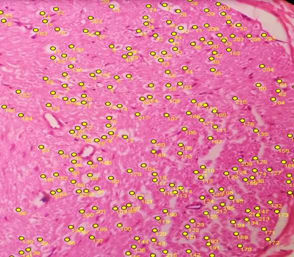
Histopathology is the science that studies the signs of disease by studying the structural and functional changes that occur in cells using certain types of dyes such as hematoxylin and eosin (H&E). Traditionally histopathological testing is carried out using semi-quantitative methods. A more advanced method is done by taking photos digitally, and then digital photos are quantified with the help of software such as ImageJ using plug-in tools. Recent advances in digital pathology require the development of more efficient computerized image analysis such as the Gaussian adaptive threshold method. This research aims to compare the calculation results of computer-assisted digitalization of histopathology using the ImageJ plugin manual method with automatic calculations using Gaussian adaptive threshold to quantify the amount of sciatic nerve cell damage in the Diabetic peripheral neuropathy (DPN) rat model. In this study, two image analysis methods were used to test their ability to measure the amount of cell damage in the sciatic nerve of normal rats using a model of diabetic neuropathy. The first method uses the ImageJ plugin manual. The second method is the Gaussian adaptive threshold method. The ImageJ plugin manual method obtained a cell abnormality value of 213 cells. Meanwhile, with the Gaussian adaptive threshold method, a value of 204 cells was obtained. The calculation results of the two methods show an insignificant difference between the methods p >0.05. This study presents a computerized morphometric image analysis method with the potential for pathology digitalization applications.
Full text article
References
2. Gurcan MN, Boucheron LE, Can A, Madabhushi A, Rajpoot NM, Yener B. Histopathological image analysis: a review. IEEE Rev Biomed Eng. 2009;2:147–71. DOI: 10.1109/rbme.2009.2034865; PMCID: PMC2910932; PMID: 20671804
3. He W, Liu T, Han Y, Ming W, Du J, Liu Y, et al. A review: The detection of cancer cells in histopathology based on machine vision. Comput Biol Med. 2022;146:105636. DOI: 10.1016/j.compbiomed.2022.105636; PMID: 35751182
4. Fulawka L, Halon A. Proliferation Index Evaluation in Breast Cancer Using ImageJ and ImmunoRatio Applications. Anticancer Res. 2016;36(8):3965–72. PMID: 27466501
5. da Silva LG, da Silva Monteiro WRS, de Aguiar Moreira TM, Rabelo MAE, de Assis EACP, de Souza GT. Fractal dimension analysis as an easy computational approach to improve breast cancer histopathological diagnosis. Appl Microsc. 2021;51(1):6. DOI: 10.1186/s42649-021-00055-w; PMCID: PMC8087740; PMID: 33929635
6. Burrai GP, Gabrieli A, Polinas M, Murgia C, Becchere MP, Demontis P, et al. Canine Mammary Tumor Histopathological Image Classification via Computer-Aided Pathology: An Available Dataset for Imaging Analysis. Animals. 2023;13(9):1563. DOI: 10.3390/ani13091563; PMCID: PMC10177203; PMID: 37174600
7. Martin B, Banner BM, Schäfer EM, Mayr P, Anthuber M, Schenkirsch G, et al. Tumor proportion in colon cancer: results from a semiautomatic image analysis approach. Virchows Arch. 2020;477(2):185–93. DOI: 10.1007/s00428-020-02764-1; PMCID: PMC7985049; PMID: 32076815
8. Wang KS, Yu G, Xu C, Meng XH, Zhou J, Zheng C, et al. Accurate diagnosis of colorectal cancer based on histopathology images using artificial intelligence. BMC Med. 2021;19(1):76. DOI: 10.1186/s12916-021-01942-5; PMCID: PMC7986569; PMID: 33752648
9. Bao J, Walliander M, Kovács F, Nagaraj AS, Hemmes A, Sarhadi VK, et al. Spa-RQ: an Image Analysis Tool to Visualise and Quantify Spatial Phenotypes Applied to Non-Small Cell Lung Cancer. Sci Rep. 2019;9(1):17613. DOI: 10.1038/s41598-019-54038-9; PMCID: PMC6879493; PMID: 31772293
10. Nagpal K, Foote D, Tan F, Liu Y, Chen PHC, Steiner DF, Manoj N, et al. Development and Validation of a Deep Learning Algorithm for Gleason Grading of Prostate Cancer from Biopsy Specimens. JAMA Oncol. 2020;6(9):1372-80. DOI: 10.1001/jamaoncol.2020.2485; PMCID: PMC7378872; PMID: 32701148
11. Kuiava VA, Kuiava EL, Chielle EO, Bittencourt FM De. Artificial intelligence algorithm for the histopathological diagnosis of skin cancer. Clin Biomed Res. 2020;40(4):218–22.
12. Li T, Xie P, Liu J, Chen M, Zhao S, Kang W, et al. Automated Diagnosis and Localization of Melanoma from Skin Histopathology Slides Using Deep Learning: A Multicenter Study. J Healthc Eng. 2021;2021:5972962. DOI: 10.1155/2021/5972962; PMCID: PMC8564171; PMID: 34745503
13. Raafat KM, El-Zahaby SA. Niosomes of active Fumaria officinalis phytochemicals: Antidiabetic, antineuropathic, anti-inflammatory, and possible mechanisms of action. Chinese Med. 2020;15:40. DOI: 10.1186/s13020-020-00321-1; PMCID: PMC7195756; PMID: 32377229
14. Shinouchi R, Shibata K, Hashimoto T, Jono S, Hasumi K, Nobe K. SMTP-44D improves diabetic neuropathy symptoms in mice through its antioxidant and anti-inflammatory activities. Pharmacol Res Perspect. 2020;8(6):e00648. DOI: 10.1002/prp2.648; PMCID: PMC7677968; PMID: 33215875
15. Sameni H, Panahi M. The Effect of Co-administration of 4-Methylcatechol and Progesterone on Sciatic Nerve Function and Neurohistological Alterations in Streptozotocin-Induced Diabetic Neuropathy in Rats. Cell J. 2011;13(1):31-8. PMCID: PMC3652538; PMID: 23671825
16. Moscalu M, Moscalu R, Dascălu CG, Țarcă V, Cojocaru E, Costin IM, et al. Histopathological Images Analysis and Predictive Modeling Implemented in Digital Pathology-Current Affairs and Perspectives. Diagnostics. 2023;13(14):2379. DOI: 10.3390/diagnostics13142379; PMCID: PMC10378281; PMID: 37510122
17. Khan A, Shal B, Khan AU, Ullah R, Baig MW, Ul Haq I, et al. Suppression of TRPV1/TRPM8/P2Y Nociceptors by Withametelin via Downregulating MAPK Signaling in Mouse Model of Vincristine-Induced Neuropathic Pain. Int J Mol Sci. 2021;22(11):6084. DOI: 10.3390/ijms22116084; PMCID: PMC8200233; PMID: 34199936
18. Labno C. Two Ways to Count Cells with ImageJ. Chicago (IL): Integrated Light Microscopy Core University of Chicago; 2014. p. 1–5. Available from: https://cpb-us-w2.wpmucdn.com/voices.uchicago.edu/dist/c/2275/files/2020/01/cell_counting_automated_and_manual.pdf
19. Lutnick B, Ramon AJ, Ginley B, Csiszer C, Kim A, Flament I, et al. Accelerating pharmaceutical R&D with a user-friendly AI system for histopathology image analysis. J Pathol Inform. 2023;14:100337. DOI: 10.1016/j.jpi.2023.100337; PMCID: PMC10582575; PMID: 37860714
20. Veta M, Pluim JPW, van Diest PJ, Viergever MA. Breast cancer histopathology image analysis: A review. IEEE Trans Biomed Eng. 2014;61(5):1400–11. DOI: 10.1109/tbme.2014.2303852; PMID: 24759275
21. Raghavan V, Rao KR. An ImageJ Based Semi-Automated Morphometric Assessment of Nuclei in Oncopathology. Int J Sci Study. 2015;3(7):189–94. DOI: 10.17354/ijss/2015/475
22. Aeffner F, Zarella MD, Buchbinder N, Bui MM, Goodman MR, Hartman DJ, et al. Introduction to Digital Image Analysis in Whole-slide Imaging: A White Paper from the Digital Pathology Association. J Pathol Inform. 2019;10:9. DOI: 10.4103/jpi.jpi_82_18; PMCID: PMC6437786; PMID: 30984469
23. Herbert A. Single Molecule Light Microscopy ImageJ Plugins. East Sussex (UK): University of Sussex; 2014. p. 1–156. Available from: http://www.sussex.ac.uk/gdsc/intranet/pdfs/SMLM.pdf
24. Anggraeni DT. Perbaikan Citra Dokumen Hasil Pindai Menggunakan Metode Simple, Adaptive-Gaussian, dan Otsu Binarization Thresholding. EXPERT J Manajemen Sistem Informasi Teknologi. 2021;11(2):71-7. DOI: 10.36448/expert.v11i2.2170
25. Korzynska A, Roszkowiak L, Lopez C, Bosch R, Witkowski L, Lejeune M. Validation of various adaptive threshold methods of segmentation applied to follicular lymphoma digital images stained with 3,3'-Diaminobenzidine&Haematoxylin. Diagn Pathol. 2013;8:48. DOI: 10.1186/1746-1596-8-48; PMCID: PMC3656801; PMID: 23531405
26. Rehman NA, Haroon F. Adaptive Gaussian and Double Thresholding for Contour Detection and Character Recognition of Two-Dimensional Area Using Computer Vision. Eng Proc. 2023;32(1):23. DOI: 10.3390/engproc2023032023
27. Kowal M, Filipczuk P, Obuchowicz A, Korbicz J. Computer-aided diagnosis of breast cancer based on fine needle biopsy microscopic images. Comput Biol Med. 2013;43(10):1563–72. DOI: 10.1016/j.compbiomed.2013.08.003; PMID: 24034748
28. Das A, Nair MS, Peter SD. Computer-Aided Histopathological Image Analysis Techniques for Automated Nuclear Atypia Scoring of Breast Cancer: a Review. J Digit Imaging. 2020;33(5):1091–121. DOI: 10.1007/s10278-019-00295-z; PMCID: PMC7573034; PMID: 31989390
29. Suarez-Arnedo A, Figueroa FT, Clavijo C, Arbeláez P, Cruz JC, Muñoz-Camargo C. An image J plugin for the high throughput image analysis of in vitro scratch wound healing assays. PLoS One. 2020;15(7):e0232565. DOI: 10.1371/journal.pone.0232565; PMCID: PMC7386569; PMID: 32722676
30. Chan HP, Hadjiiski LM, Samala RK. Computer-aided diagnosis in the era of deep learning. Med Phys. 2020;47(5):e218-27. DOI: 10.1002/mp.13764; PMCID: PMC7293164; PMID: 32418340
31. Brixtel R, Bougleux S, Lézoray O, Caillot Y, Lemoine B, Fontaine M, et al. Whole Slide Image Quality in Digital Pathology: Review and Perspectives. IEEE Access. 2022;10:131005-35. DOI: 10.1109/ACCESS.2022.3227437
Authors
Copyright (c) 2024 Indah Tri Lestari, Kusnandar Anggadiredja, Afrillia Nuryanti Garmana, Sevi Nurafni

This work is licensed under a Creative Commons Attribution-ShareAlike 4.0 International License.
This work is licensed under a Creative Commons Attribution-ShareAlike 4.0 International License.
Authors continue to retain the copyright to the article if the article is published in the Borneo Journal of Pharmacy. They will also retain the publishing rights to the article without any restrictions.
Authors who publish in this journal agree to the following terms:
- Any article on the copyright is retained by the author(s).
- The author grants the journal the right of first publication with the work simultaneously licensed under a Creative Commons Attribution License that allows others to share work with an acknowledgment of the work authors and initial publications in this journal.
- Authors can enter into separate, additional contractual arrangements for the non-exclusive distribution of published articles (e.g., post-institutional repository) or publish them in a book, with acknowledgment of their initial publication in this journal.
- Authors are permitted and encouraged to post their work online (e.g., in institutional repositories or on their websites) prior to and during the submission process. This can lead to productive exchanges and earlier and greater citations of published work.
- The article and any associated published material are distributed under the Creative Commons Attribution-ShareAlike 4.0 International License.
Article Details


