Campylobacter Species, Microbiological Source Tracking and Risk Assessment of Bacterial pathogens Array

Article Sidebar
-
campylobacter, campylobacteriosis, microbial risk assessment, source attribution, bacterial pathogens
Abstract
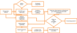
Campylobacter species continue to remain critical pathogens of public health interest. They are responsible for approximately 500 million cases of gastroenteritis per year worldwide. Infection occurs through the consumption of contaminated food and water. Microbial risk assessment and source tracking are crucial epidemiological strategies to monitor the outbreak of campylobacteriosis effectively. Various methods have been proposed for microbial source tracking and risk assessment, most of which rely on conventional microbiological techniques such as detecting fecal indicator organisms and other novel microbial source tracking methods, including library-dependent microbial source tracking and library-independent source tracking approaches. However, both the traditional and novel methods have their setbacks. For example, while the conventional techniques are associated with a poor correlation between indicator organism and pathogen presence, on the other hand, it is impractical to interpret qPCR-generated markers to establish the exact human health risks even though it can give information regarding the potential source and relative human risk. Therefore, this article provides up-to-date information on campylobacteriosis, various approaches for source attribution, and risk assessment of bacterial pathogens, including next-generation sequencing approaches such as shotgun metagenomics, which effectively answer the questions of potential pathogens are there and in what quantities.
Full text article
References
2. Aslam B, Wang W, Arshad MI, Khurshid M, Muzammil S, Rasool MH, et al. Antibiotic resistance: a rundown of a global crisis. Infect Drug Resist. 2018;11:1645-58. doi:10.2147/idr.s173867
3. Manyi-Loh C, Mamphweli S, Meyer E, Okoh A. Antibiotic Use in Agriculture and Its Consequential Resistance in Environmental Sources: Potential Public Health Implications. Molecules. 2018;23(4):795. doi:10.3390/molecules23040795
4. Facciolà A, Riso R, Avventuroso E, Visalli G, Delia SA, Laganà P. Campylobacter: from microbiology to prevention. J Prev Med Hyg. 2017;58(2):E79-92.
5. Rodrigues C, Cunha MÂ. Assessment of the microbiological quality of recreational waters: indicators and methods. Euro-Mediterr J Environ Integr. 2017;2:25. doi:10.1007/s41207-017-0035-8
6. Li E, Saleem F, Edge TA, Schellhorn HE. Biological Indicators for Fecal Pollution Detection and Source Tracking: A Review. Processes. 2021;9(11):2058. doi:10.3390/pr9112058
7. Teixeira P, Dias D, Costa S, Brown B, Silva S, Valério E. Bacteroides spp. and traditional fecal indicator bacteria in water quality assessment - An integrated approach for hydric resources management in urban centers. J Environ Manage. 2020;271:110989. doi:10.1016/j.jenvman.2020.110989
8. Ahmed W, Hamilton K, Toze S, Cook S, Page D. A review on microbial contaminants in stormwater runoff and outfalls: Potential health risks and mitigation strategies. Sci Total Environ. 2019;692:1304-21. doi:10.1016/j.scitotenv.2019.07.055
9. Edge TA, Hill S, Seto P, Marsalek J. Library-dependent and library-independent microbial source tracking to identify spatial variation in faecal contamination sources along a Lake Ontario beach (Ontario, Canada). Water Sci Technol. 2010;62(3):719-27. doi:10.2166/wst.2010.335
10. Vadde KK, McCarthy AJ, Rong R, Sekar R. Quantification of Microbial Source Tracking and Pathogenic Bacterial Markers in Water and Sediments of Tiaoxi River (Taihu Watershed). Front Microbiol. 2019;10:699. doi:10.3389/fmicb.2019.00699
11. Schuppler M, Lötzsch K, Waidmann M, Autenrieth IB. An abundance of Escherichia coli is harbored by the mucosa-associated bacterial flora of interleukin-2-deficient mice. Infect Immun. 2004;72(4):1983-90. doi:10.1128/iai.72.4.1983-1990.2004
12. Liu R, Chiang MHY, Lun CHI, Qian PY, Lau SCK. Host-specific 16S rRNA gene markers of Bacteroidales for source tracking of fecal pollution in the subtropical coastal seawater of Hong Kong. Water Res. 2010;44(20):6164-74. doi:10.1016/j.watres.2010.07.035
13. Green HC, Dick LK, Gilpin B, Samadpour M, Field KG. Genetic markers for rapid PCR-based identification of gull, Canada goose, duck, and chicken fecal contamination in water. Appl Environ Microbiol. 2012;78(2):503–10. doi:10.1128/aem.05734-11
14. Odagiri M, Schriewer A, Hanley K, Wuertz S, Misra PR, Panigrahi P, et al. Validation of Bacteroidales quantitative PCR assays targeting human and animal fecal contamination in the public and domestic domains in India. Sci Total Environ. 2015;502:462–70. doi:10.1016/j.scitotenv.2014.09.040
15. Boehm AB, Wang D, Ercumen A, She M, Harris AR, Shanks OC, et al. Occurrence of host-associated fecal markers on child hands, household soil, and drinking water in rural Bangladeshi households. Environ Sci Technol Lett. 2016;3(11):393–8. doi:10.1021/acs.estlett.6b00382
16. Shanks OC, Atikovic E, Blackwood AD, Lu J, Noble RT, Domingo JS, et al. Quantitative PCR for detection and enumeration of genetic markers of bovine fecal pollution. Appl Environ Microbiol. 2008;74(3):745-52. doi:10.1128/aem.01843-07
17. Kraemer SA, Ramachandran A, Perron GG. Antibiotic Pollution in the Environment: From Microbial Ecology to Public Policy. Microorganisms. 2019;7(6):180. doi:10.3390/microorganisms7060180
18. Choi Y, Oda E, Waldman O, Sajda T, Beck C, Oh I. Next-Generation Sequencing for Pathogen Identification in Infected Foot Ulcers. Foot Ankle Orthop. 2021;6(3):24730114211026933. doi:10.1177/24730114211026933
19. Couto N, Schuele L, Raangs EC, Machado MP, Mendes CI, Jesus TF, et al. Critical steps in clinical shotgun metagenomics for the concomitant detection and typing of microbial pathogens. Sci Rep. 2018;8(1):13767. doi:10.1038/s41598-018-31873-w
20. Buytaers FE, Saltykova A, Denayer S, Verhaegen B, Vanneste K, Roosens NHC, et al. A Practical Method to Implement Strain-Level Metagenomics-Based Foodborne Outbreak Investigation and Source Tracking in Routine.Microorganisms. 2020;8(8):1191. doi:10.3390/microorganisms8081191
21. Zhang L, Chen F, Zeng Z, Xu M, Sun F, Yang L, et al. Advances in Metagenomics and Its Application in Environmental Microorganisms. Front Microbiol. 2021;12:766364. doi:10.3389/fmicb.2021.766364
22. Donia MS, Cimermancic P, Schulze CJ, Brown LCW, Martin J, Mitreva M, et al. A systematic analysis of biosynthetic gene clusters in the human microbiome reveals a common family of antibiotics. Cell. 2014;158(6):1402–14. doi:10.1016/j.cell.2014.08.032
23. Norman JM, Handley SA, Baldridge MT, Droit L, Liu CY, Keller BC, et al. Disease-specific alterations in the enteric virome in inflammatory bowel disease. Cell. 2015;160(3):447–60. doi:10.1016/j.cell.2015.01.002
24. Loman NJ, Constantinidou C, Christner M, Rohde H, Chan JZM, Quick J, et al. A culture-independent sequence-based metagenomics approach to the investigation of an outbreak of Shiga-toxigenic Escherichia coli O104:H4. JAMA. 2013;309(14):1502–10. doi:10.1001/jama.2013.3231
25. Cocolin L, Mataragas M, Bourdichon F, Doulgeraki A, Pilet MF, Jagadeesan B, et al. Next generation microbiological risk assessment meta-omics : The next need for integration. Int J Food Microbiol. 2018;287:10-7. doi:10.1016/j.ijfoodmicro.2017.11.008
26. Veron M, Chatelain R. Taxonomic study of the genus Campylobacter Sebald and Veron and designation of the neotype strain for the type species, Campylobacter fetus (Smith and Taylor) Sebald and Veron. Int J Syst Evol Microbiol. 1973;23(2):122–34. doi:10.1099/00207713-23-2-122
27. Whitehouse CA, Zhao S, Tate H. Antimicrobial Resistance in Campylobacter Species : Mechanisms and Genomic Epidemiology. Adv Appl Microbiol. 2018;103:1-47. doi:10.1016/bs.aambs.2018.01.001
28. Dasti JI, Tareen AM, Lugert R, Zautner AE, Gross U. Campylobacter jejuni: A brief overview on pathogenicity-associated factors and disease-mediating mechanisms. Int J Med Microbiol. 2010;300(4):205-11. doi:10.1016/j.ijmm.2009.07.002
29. Iraola G, Pérez R, Naya H, Paolicchi F, Pastor E, Valenzuela S, et al. Genomic Evidence for the Emergence and Evolution of Pathogenicity and Niche Preferences in the Genus Campylobacter. Genome Biol Evol. 2014;6(9):2392–405. doi:10.1093/gbe/evu195
30. Hatanaka N, Shimizu A, Somroop S, Li Y, Asakura M, Nagita A, et al. High prevalence of Campylobacter ureolyticus in stool specimens of children with diarrhea in Japan. Jpn J Infect Dis. 2017;70(4):455–7. doi:10.7883/yoken.jjid.2016.428
31. Chen Y, Mukherjee S, Hoffmann M, Kotewicz ML, Young S, Abbott J, et al. Whole-genome sequencing of gentamicin-resistant Campylobacter coli isolated from U.S. retail meats reveals novel plasmid-mediated aminoglycoside resistance genes. Antimicrob Agents Chemother. 2013;57(11):5398–405. doi:10.1128/aac.00669-13
32. Gibreel A, Wetsch NM, Taylor DE. Contribution of the CmeABC efflux pump to macrolide and tetracycline resistance in Campylobacter jejuni. Antimicrob Agents Chemother. 2007;51(9):3212–16. doi:10.1128/aac.01592-06
33. Shobo CO, Bester LA, Baijnath S, Somboro AM, Peer AKCC, Essack SY. Original Article Antibiotic resistance profiles of Campylobacter species in the South Africa private health care sector. J Infect Dev Ctries. 2016;10(11):1214-21. doi:10.3855/jidc.8165
34. Acke E. Campylobacteriosis in dogs and cats: a review. N Z Vet J. 2018;66(5):221-8. doi:10.1080/00480169.2018.1475268
35. Mourkas E, Florez-Cuadrado D, Pascoe B, Calland JK, Bayliss SC, Mageiros L, et al. Gene pool transmission of multidrug resistance among Campylobacter from livestock, sewage and human disease. Environ Microbiol. 2019;21(12):4597-613. doi:10.1111/1462-2920.14760
36. Tyson GH, McDermott PF, Li C, Chen Y, Tadesse DA, Mukherjee S, et al. WGS accurately predicts antimicrobial resistance in Escherichia coli. J Antimicrob Chemother. 2015;70(10):2763–9. doi:10.1093/jac/dkv186
37. Rosner BM, Schielke A, Didelot X, Kops F, Breidenbach J, Willrich N, et al. A combined case-control and molecular source attribution study of human Campylobacter infections in Germany, 2011–2014. Sci Rep. 2017;7(1):5139. doi:10.1038/s41598-017-05227-x
38. Chukwu MO, Luther A, Abia K, Ubomba-jaswa E, Obi L, Dewar JB. Characterization and Phylogenetic Analysis of Campylobacter Species Isolated from Paediatric Stool and Water Samples in the Northwest Province, South Africa. Int J Environ Res Public Health. 2019;16(12):2205. doi:10.3390/ijerph16122205
39. Bourke B, Chan VL, Sherman P. Campylobacter upsaliensis: Waiting in the wings. Clin Microbiol Rev. 1998;11(3):440–9. doi:10.1128/cmr.11.3.440
40. Abril C, Brodard I, Perreten V. Located within a Transferable Pathogenicity Island in Campylobacter fetus subsp. fetus. Antimicrob Agents Chemother. 2010;54(7):3052-5. doi:10.1128/aac.00304-10
41. Wagenaar JA, van Bergen MA, Blaser MJ, Tauxe RV, Newell DG, van Putten JP. Campylobacter fetus infections in humans: Exposure and disease. Clin Infect Dis. 2014;58(11):1579–86. doi:10.1093/cid/ciu085
42. Roe DE, Weinberg A, Roberts MC. Mobile rRNA methylase genes in Campylobacter (Wolinella) rectus. J Antimicrob Chemother. 1995;36(4):738–40. doi:10.1093/jac/36.4.738
43. Mahlen SD, Clarridge JE. Oral Abscess Caused by Campylobacter rectus: Case Report and Literature Review ᰔ. J Clin Microbiol. 2009;47(3):848-51. doi:10.1128/jcm.01590-08
44. Man SM. The clinical importance of emerging Campylobacter species. Nat Rev Gastroenterol Hepatol. 2011;8(12):669-85. doi:10.1038/nrgastro.2011.191
45. Laatu M, Rautelin H, Hänninen ML. Susceptibility of Campylobacter hyointestinalis subsp. hyointestinalis to antimicrobial agents and characterization of quinolone-resistant strains. J Antimicrob Chemother. 2005;55(2):182-7. doi:10.1093/jac/dkh537
46. Iovine NM. Resistance mechanisms in Campylobacter jejuni. Virulence. 2013;4(3):230-40. doi:10.4161/viru.23753
47. Taylor DE, Courvalin P. Mechanisms of antibiotic resistance in Campylobacter species. Antimicrob Agents Chemother. 1988;32(8):1107–12. doi:10.1128/aac.32.8.1107
48. Wieczorek K, Osek J. Antimicrobial resistance mechanisms among Campylobacter. Biomed Res Int. 2013;2013:340605. doi:10.1155/2013/340605
49. Munita JM, Arias CA. Mechanisms of Antibiotic Resistance. Microbiol Spectr. 2016;4(2):10.1128/microbiolspec.VMBF-0016-2015. doi:10.1128/microbiolspec.vmbf-0016-2015
50. Hamilton AJ, Stagnitti F, Premier R, Boland AM, Hale G. Quantitative microbial risk assessment models for consumption of raw vegetables irrigated with reclaimed water. Appl Environ Microbiol. 2006;72(5):3284-90. doi:10.1128/aem.72.5.3284-3290.2006
51. Eisenberg JNS, Lei X, Hubbard AH, Brookhart MA, Colford JM. The role of disease transmission and conferred immunity in outbreaks: Analysis of the 1993 Cryptosporidium outbreak in Milwaukee, Wisconsin. Am J Epidemiol. 2005;161(1):62–72. doi:10.1093/aje/kwi005
52. Eisenberg JNS, Brookhart MA, Rice G, Brown M, Colford JM. Disease transmission models for public health decision making: Analysis of epidemic and endemic conditions caused by waterborne pathogens. Environ Health Perspect. 2002;110(8):783–90. doi:10.1289/ehp.02110783
53. Brouwer AF, Masters NB, Eisenberg JNS. Quantitative Microbial Risk Assessment and Infectious Disease Transmission Modeling of Waterborne Enteric Pathogen. Curr Environ Health Rep. 2019;5(2):293-304. doi:10.1007/s40572-018-0196-x
54. Germovsek E, Barker CI, Sharland M. What do I need to know about aminoglycoside antibiotics? Arch Dis Child Educ Pract Ed. 2017;102(2):89-93. doi:10.1136/archdischild-2015-309069
55. Schwarz S, Shen J, Kadlec K, Wang Y, Michael GB, Feßler AT, et al. Lincosamides, Streptogramins, Phenicols, and Pleuromutilins: Mode of Action and Mechanisms of Resistance. Cold Spring Harb Perspect Med. 2016;6(11):a027037. doi:10.1101/cshperspect.a027037
56. Roberts MC. Update on macrolide-lincosamide-streptogramin, ketolide, and oxazolidinone resistance genes. FEMS Microbiol Lett. 2008;282(2):147-59. doi:10.1111/j.1574-6968.2008.01145.x
57. Pham TDM, Ziora ZM, Blaskovich MAT. Quinolone antibiotics. Medchemcomm. 2019;10(10):1719-39. doi:10.1039/c9md00120d
58. Pulicharla R, Hegde K, Brar SK, Surampalli RY. Tetracyclines metal complexation: Significance and fate of mutual existence in the environment. Environ Pollut. 2017;221:1-14. doi:10.1016/j.envpol.2016.12.017
59. Abdi-Hachesoo B, Khoshbakht R, Sharifiyazdi H, Tabatabaei M, Hosseinzadeh S, Asasi K. Tetracycline Resistance Genes in Campylobacter jejuni and C. coli Isolated from Poultry Carcasses. Jundishapur J Microbiol. 2014;7(9):e12129. doi:10.5812/jjm.12129
60. Bush K, Bradford PA. β-Lactams and β-Lactamase Inhibitors: An Overview. Cold Spring Harb Perspect Med. 2016;6(8):a025247. doi: https://doi.org/10.1101/cshperspect.a025247
61. Chandrasekaran S, Jiang SC. A dose response model for quantifying the infection risk of antibiotic-resistant bacteria. Sci Rep. 2019;9(1):17093. doi:10.1038/s41598-019-52947-3
62. Haas CN. Microbial dose response modeling: past, present, and future. Environ Sci Technol. 2015;49(3):1245-59. doi:10.1021/es504422q
63. Nauta MJ. Modelling bacterial growth in quantitative microbiological risk assessment: is it possible? Int J Food Microbiol. 2002;73(2-3):297-304. doi:10.1016/s0168-1605(01)00664-x
64. Ahmed Z, Mohamed K, Zeeshan S, Dong X. Artificial intelligence with multi-functional machine learning platform development for better healthcare and precision medicine. Database. 2020;2020:baaa010. doi:10.1093/database/baaa010
65. Weir MH, Mitchell J, Flynn W, Pope JM. Development of a microbial dose response visualization and modelling application for QMRA modelers and educators. Environ Model Softw. 2017;88:74-83. doi:10.1016/j.envsoft.2016.11.011
66. Schijven JF, Teunis PFM, Rutjes SA, Bouwknegt M, Husman AMdR. QMRAspot: a tool for Quantitative Microbial Risk Assessment from surface water to potable water. Water Res. 2011;45(17):5564-76. doi:10.1016/j.watres.2011.08.024
67. Chen Y, Dennis SB, Hartnett E, Paoli G, Pouillot R, Ruthman T, et al. FDA-iRISK--a comparative risk assessment system for evaluating and ranking food-hazard pairs: case studies on microbial hazards. J Food Prot. 2013;76(3):376-85. doi:10.4315/0362-028x.jfp-12-372
68. Ercolini D. High-throughput sequencing and metagenomics: moving forward in the culture-independent analysis of food microbial ecology. Appl Environ Microbiol. 2013;79(10):3148-55. doi:10.1128/aem.00256-13
69. Sharpton TJ. An introduction to the analysis of shotgun metagenomic data. Front Plant Sci. 2014;5:209. doi:10.3389/fpls.2014.00209
70. Tengh F, Nair SSD, Zhu P, Li S, Huang S, Li X, et al. Impact of DNA extraction method and targeted 16S-rRNA hypervariable region on oral microbiota profiling. Sci Rep. 2018;8(1):16321. doi:10.1038/s41598-018-34294-x
71. Durazzi F, Sala C, Castellani G, Manfreda G, Remondini D, De Cesare A. Comparison between 16S rRNA and shotgun sequencing data for the taxonomic characterization of the gut microbiota. Sci Rep. 2021;11(1):3030. doi:10.1038/s41598-021-82726-y
72. Vecherskii MV, Semenov MV, Lisenkova AA, Stepankov AA. Metagenomics: A New Direction in Ecology. Biol Bull Russ Acad Sci. 2021;48:S107-17. doi:10.1134/S1062359022010150
73. Dai D, Rhoads WJ, Edwards MA, Pruden A. Shotgun Metagenomics Reveals Taxonomic and Functional Shifts in Hot Water Microbiome Due to Temperature Setting and Stagnation. Front Microbiol. 2018;9:2695. doi:10.3389/fmicb.2018.02695
74. Chen H, Li Y, Sun W, Song L, Zuo R, Teng Y. Characterization and source identi fi cation of antibiotic resistance genes in the sediments of an interconnected river-lake system. Environ Int. 2020;137:105538. doi:10.1016/j.envint.2020.105538
75. Mohiuddin MM, Salama Y, Schellhorn HE, Golding GB. Shotgun metagenomic sequencing reveals freshwater beach sands as reservoir of bacterial pathogens. Water Res. 2017;115:360-9. doi:10.1016/j.watres.2017.02.057
76. Pérez-Cobas AE, Gomez-Valero L, Buchrieser C. Metagenomic approaches in microbial ecology: an update on whole-genome and marker gene sequencing analyses. Microb Genom. 2020;6(8):mgen000409. doi:10.1099/mgen.0.000409
77. Clarridge JE. Impact of 16S rRNA gene sequence analysis for identification of bacteria on clinical microbiology and infectious diseases. Clin Microbiol Rev. 2004;17(4):840-62. doi:10.1128/cmr.17.4.840-862.2004
78. Jovel J, Patterson J, Wang W, Hotte N, O’Keefe S, Mitchel T, et al. Characterization of the Gut Microbiome Using 16S or Shotgun Metagenomics. Front Microbiol. 2016;7:459. doi:10.3389/fmicb.2016.00459
79. Stewart MP, Langer R, Jensen KF. Intracellular Delivery by Membrane Disruption: Mechanisms, Strategies, and Concepts. Chem Rev. 2018;118(16):7409-531. doi:10.1021/acs.chemrev.7b00678
80. Awasthi MK, Ravindran B, Sarsaiya S, Chen H, Wainaina S, Singh E, et al. Metagenomics for taxonomy profiling: tools and approaches. Bioengineered. 2020;11(1):356-74. doi:10.1080/21655979.2020.1736238
81. Grützke J, Gwida M, Deneke C, Brendebach H, Projahn M, Schattschneider A, et al. Direct identification and molecular characterization of zoonotic hazards in raw milk by metagenomics using Brucella as a model pathogen. Microb Genom. 2021;7(5):000552. doi:10.1099/mgen.0.000552
82. EFSA Panel on Biological Hazards (EFSA BIOHAZ Panel), Koutsoumanis K, Allende A, Alvarez-Ordóñez A, Bolton D, Bover-Cid S, et al. Whole genome sequencing and metagenomics for outbreak investigation, source attribution and risk assessment of food-borne microorganisms. EFSA J. 2019;17(12):e05898. doi:10.2903/j.efsa.2019.5898
83. Priyanka B, Patil RK, Dwarakanath S. A review on detection methods used for foodborne pathogens. Indian J Med Res. 2016;144(3):327-38. doi:10.4103/0971-5916.198677
84. Uelze L, Grützke J, Borowiak M, Hammerl JA, Juraschek K, Deneke C, et al. Typing methods based on whole genome sequencing data. One Health Outlook. 2020;2:3. doi:10.1186/s42522-020-0010-1
85. Underwood AP, Dallman T, Thomson NR, Williams M, Harker K, Perry N, et al. Public health value of next-generation DNA sequencing of enterohemorrhagic Escherichia coli isolates from an outbreak. J Clin Microbiol. 2013;51(1):232–7. doi:10.1128/jcm.01696-12
86. Hoffmann M, Luo Y, Monday SR, Gonzalez-Escalona N, Ottesen AR, Muruvanda T, et al. Tracing origins of the Salmonella Bareilly strain causing a food-borne outbreak in the united states. J Infect Dis. 2016;213(4):502–8. doi:10.1093/infdis/jiv297
87. Kleta S, Hammerl JA, Dieckmann R, Malorny B, Borowiak M, Halbedel S, et al. Molecular tracing to find source of protracted invasive listeriosis outbreak, southern Germany, 2012–2016. Emerg Infect Dis. 2017;23(10):1680–3. doi:10.3201/eid2310.161623
88. Allard MW, Strain E, Melka D, Buning K, Musser SM, Brown EW, et al. The practical value of food pathogen traceability through building a wholegenome sequencing network and database. J Clin Microbiol. 2016;54(8):1975–83. doi:10.1128/jcm.00081-16
89. Rantsiou K, Kathariou S, Winkler A, Skandamis P, Saint-Cyr MJ, Rouzeau-Szynalski K, et al. Next generation microbiological risk assessment: opportunities of whole genome sequencing (WGS) for foodborne pathogen surveillance, source tracking and risk assessment. Int J Food Microbiol. 2018;287:3-9. doi:10.1016/j.ijfoodmicro.2017.11.007
90. Besser JM, Carleton HA, Trees E, Stroika SG, Hise K, Wise M, et al. Interpretation of Whole-Genome Sequencing for Enteric Disease Surveillance and Outbreak Investigation. Foodborne Pathog Dis. 2019;16(7):504-12. doi:10.1089/fpd.2019.2650
91. Stein RA, Chirilã M. Routes of Transmission in the Food Chain. Foodborne Dis. 2017;65-13. doi:10.1016/B978-0-12-385007-2.00003-6
92. Afshinnekoo E, Chou C, Alexander N, Ahsanuddin S, Schuetz AN, Mason CE. Precision Metagenomics: Rapid Metagenomic Analyses for Infectious Disease Diagnostics and Public Health Surveillance. J Biomol Tech. 2017;28(1):40-5. doi:10.7171/jbt.17-2801-007
93. Buytaers FE, Saltykova A, Mattheus W, Verhaegen B, Roosens NHC, Vanneste K, et al. Application of a strain-level shotgun metagenomics approach on food samples: resolution of the source of a Salmonella food-borne outbreak. Microb Genome. 2021;7(4):000547. doi:10.1099/mgen.0.000547
94. Piombo E, Abdelfattah A, Droby S, Wisniewski M, Spadaro D, Schena L. Metagenomics Approaches for the Detection and Surveillance of Emerging and Recurrent Plant Pathogens. Microorganisms. 2021;9(1):188. doi:10.3390/microorganisms9010188
95. Larsson DGJ, Flach CF. Antibiotic resistance in the environment. Nat Rev Microbiol. 2022;20(5):257-69. doi:10.1038/s41579-021-00649-x
96. Höper D, Grützke J, Brinkmann A, Mossong J, Matamoros S, Ellis RJ, et al. Proficiency Testing of Metagenomics-Based Detection of Food-Borne Pathogens Using a Complex Artificial Sequencing Dataset. Front Microbiol. 2020;11:575377. doi:10.3389/fmicb.2020.575377
97. Grützke J, Malorny B, Hammerl JA, Busch A, Tausch SH, Tomaso H, et al. Fishing in the Soup – Pathogen Detection in Food Safety Using Metabarcoding and Metagenomic Sequencing. Front Microbiol. 2019;10:1805. doi:10.3389/fmicb.2019.01805
98. Bharucha T, Oeser C, Balloux F, Brown JR, Carbo EC, Charlett A, et al. STROBE-metagenomics: a STROBE extension statement to guide the reporting of metagenomics studies. Lancet Infect Dis. 2020;20(10):e251-60. doi:10.1016/s1473-3099(20)30199-7
99. Lazou TP, Gelasakis AI, Chaintoutis SC, Iossifidou EG, Dovas CI. Method-Dependent Implications in Foodborne Pathogen Quantification: The Case of Campylobacter coli Survival on Meat as Comparatively Assessed by Colony Count and Viability PCR. Front Microbiol. 2021;12:604933. doi:10.3389/fmicb.2021.604933
100. Lagier JC, Edouard S, Pagnier I, Mediannikov O, Drancourt M, Raoult D. Current and past strategies for bacterial culture in clinical microbiology. Clin Microbiol Rev. 2015;28(1):208-36. doi:10.1128/cmr.00110-14
101. Liu Y, Gilchrist A, Zhang J, Li XF. Detection of viable but nonculturable Escherichia coli O157:H7 bacteria in drinking water and river water. Appl Enciron Microbiol. 2008;74(5):1502-7. doi:10.1128/aem.02125-07
102. Schofield DA, Sharp NJ, Westwater C. Phage-based platforms for the clinical detection of human bacterial pathogens. Bacteriophage. 2012;2(2):105-283. doi:10.4161/bact.19274
103. Cangelosi G, Meschke JS. Dead or alive: molecular assessment of microbial viability. Appl Environ Microbiol. 2014;80(19):5884-91. doi:10.1128/aem.01763-14
104. Zeng D, Chen Z, Jiang Y, Xue F, Li B. Advances and Challenges in Viability Detection of Foodborne Pathogens. Front Microbiol. 2016;7:1833. doi:10.3389/fmicb.2016.01833
105. Cancino-Faure B, Fisa R, Alcover MM, Jimenez-Marco T, Riera C. Detection and Quantification of Viable and Nonviable Trypanosoma cruzi Parasites by a Propidium Monoazide Real-Time Polymerase Chain Reaction Assay. Am J Trop Med Hyg. 2016;94(6):1282-9. doi:10.4269/ajtmh.15-0693
106. van Seventer JM, Hochberg NS. Principles of Infectious Diseases: Transmission, Diagnosis, Prevention, and Control. Int Encycl Public Health. 2017;22-39. doi:10.1016/B978-0-12-803678-5.00516-6
107. Yadav SK, Akhter Y. Statistical Modeling for the Prediction of Infectious Disease Dissemination With Special Reference to COVID-19 Spread. Front Public Health. 2021;9:645405. doi:10.3389/fpubh.2021.645405
108. Siettos CI, Russo L. Mathematical modeling of infectious disease dynamics. Virulence. 2013;4(4):295-306. doi:10.4161/viru.24041
109. Cooper I, Mondal A, Antonopoulos CG. A SIR model assumption for the spread of COVID-19 in different communities. Chaos Solitons Fractals. 2020;139:110057. doi:10.1016/j.chaos.2020.110057
110. van den Driessche P. Reproduction numbers of infectious disease models. Infect Dis Model. 2017;2(3):288-303. doi:10.1016/j.idm.2017.06.002
111. Cucinotta D, Vanelli M. WHO Declares COVID-19 a Pandemic. Acta Biomed. 2020;91(1):157-60. doi:10.23750/abm.v91i1.9397
112. Liu T, Bai Y, Du M, Gao Y, Liu Y. Susceptible-Infected-Removed Mathematical Model under Deep Learning in Hospital Infection Control of Novel Coronavirus Pneumonia. J Healthc Eng. 2021;2021:1535046. doi:10.1155/2021/1535046
113. Abou-Ismail A. Compartmental Models of the COVID-19 Pandemic for Physicians and Physician-Scientists. SN Compr Clin Med. 2020;2(7):852-8. doi:10.1007/s42399-020-00330-z
114. Brauer F. Mathematical epidemiology: Past, present, and future. Infect Dis Model. 2017;2(2):113-27. doi:10.1016/j.idm.2017.02.001
115. Nikolaou M. Ziegler and Nichols meet Kermack and McKendrick: Parsimony in dynamic models for epidemiology. Comput Chem Eng. 2022;157:107615. doi:10.1016/j.compchemeng.2021.107615
116. Howard LM, Zhu Y, Griffin MR, Edwards KM, Williams JV, Gil AI, et al. Nasopharyngeal Pneumococcal Density during Asymptomatic Respiratory Virus Infection and Risk for Subsequent Acute Respiratory Illness. Emerg Infect Dis. 2019;25(11):2040-7. doi:10.3201/eid2511.190157
117. London AJ. Artificial intelligence in medicine: Overcoming or recapitulating structural challenges to improving patient care? Cell Rep Med. 2022;3(5):100622. doi:10.1016/j.xcrm.2022.100622
118. Njage PMK, Henri C, Leekitcharoenphon P, Mistou MY, Hendriksen RS, Hald T. Machine Learning Methods as a Tool for Predicting Risk of Illness Applying Next-Generation Sequencing Data. Risk Anal. 2019;39(6):1397-413. doi:10.1111/risa.13239
119. Hedge J, Rokseth B. Applications of machine learning methods for engineering risk assessment – A review. Safety Sci. 2020;122:104492. doi:10.1016/j.ssci.2019.09.015
120. Nicholls HL, John CR, Watson DS, Munroe PB, Barnes MR, Cabrera CP. Reaching the End-Game for GWAS: Machine Learning Approaches for the Prioritization of Complex Disease Loci. Front Genet. 2020;11:350 doi:10.3389/fgene.2020.00350
Authors
Copyright (c) 2022 Bashar Haruna Gulumbe, Abbas Yusuf Bazata, Musbahu Abdullahi Bagwai

This work is licensed under a Creative Commons Attribution-ShareAlike 4.0 International License.
This work is licensed under a Creative Commons Attribution-ShareAlike 4.0 International License.
Authors continue to retain the copyright to the article if the article is published in the Borneo Journal of Pharmacy. They will also retain the publishing rights to the article without any restrictions.
Authors who publish in this journal agree to the following terms:
- Any article on the copyright is retained by the author(s).
- The author grants the journal the right of first publication with the work simultaneously licensed under a Creative Commons Attribution License that allows others to share work with an acknowledgment of the work authors and initial publications in this journal.
- Authors can enter into separate, additional contractual arrangements for the non-exclusive distribution of published articles (e.g., post-institutional repository) or publish them in a book, with acknowledgment of their initial publication in this journal.
- Authors are permitted and encouraged to post their work online (e.g., in institutional repositories or on their websites) prior to and during the submission process. This can lead to productive exchanges and earlier and greater citations of published work.
- The article and any associated published material are distributed under the Creative Commons Attribution-ShareAlike 4.0 International License.
Article Details


